Preprosthetic surgery is surgery that will prepare the mouth so that one may be able to have dentures, partials, and/or some other prosthesis fabricated and placed comfortably in the mouth.
Some patients require minor oral surgical procedures in order to ensure the maximum level of retention and comfort of the dentures. A denture sits on the bone ridge, so it is very important that the bone is the proper shape. If a tooth needs to be extracted, the underlying bone might be left sharp and uneven. For the best fit of a denture, the bone might need to be smoothed out or reshaped. Occasionally, excess bone, called tori or exostoses, would need to be removed prior to denture insertion.
One or more of the following procedures might need to be performed in order to prepare the mouth for a denture:
.
.
.
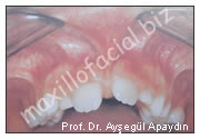
Ridge Augmentation – increasing bone in the alveolar ridge (Figure 1)
The alveolar ridge is the horseshoe shaped ridge of bone that all the teeth are rooted in. Alveolar bone loss occurs as the teeth are removed due to loss of stimulation of the bone. When replacing the teeth with an implant, a bridge or a denture it may be necessary to replace or augment the bone. This is done by bone grafting.
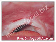
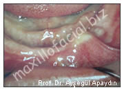
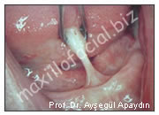
Vestibuloplasty – increasing the vestibular depth (Figure 2)
Loss of alveolar bone as mentioned above makes dentures not fit well. The denture needs a certain amount of ridge height to give it retention. Besides making the ridge bigger by adding bone one can also move the unattached (cheek tissues) gingiva away from the ridge. The vestibule is that part between the cheek and gums. Plasty of the vestibule can be done by various methods, the most reliable of which is grafting tissue obtained from another area. This could be obtained from the roof of mouth or the skin. The patient can gain tissue where it’s needed and remove unwanted tissue all at once. The roof of the mouth (hard palate) is an excellent source of attached gingiva (gums). The tissue is the same type and thickness of the needed tissue and its source, the palate, is a renewable source. A graft taken from the palate leaves a patch which will completely grow back and can be used again in the future. The drawback to this procedure is that it is fairly painful. Of course, prescription for strong pain medicines is provided and a plastic shield is made to fit over the palate to protect it and decrease the pain.
.
.
.
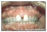
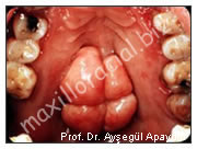
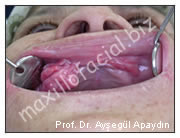
Alveoloplasty - bone smoothing and reshaping (Figure 3,4)
Alveoloplasty is simply reshaping the alveolar bone. This is the horseshoe shaped bone teeth are rooted in. The alveolar bone sits on the basal bone of the maxilla (upper jaw) and mandible (lower jaw). Any time a tooth is removed alveoloplasty may be required to smooth rough or sharp edges or reduce protruding bone. When alveoloplasty is performed, it is done in the most conservative manner possible. Preservation of the bone is important because it naturally diminishes over time. When preparing the mouth for dentures the alveolar bone may need to be shaped so the dentures can fit over it. If overhanging bone creates an undercut the bone must be reshaped.
.
.
.
Torectomy (Tori removal) - removal of excess bone (Figure 5,6)
Tori are bony "bumps" or protuberances that occur on the tongue side of lower jaw or in palate (roof of mouth). These areas of bone are generally considered to be "normal" bone and do not represent a pathologic process. One may or may not be aware of their presence due to the fact that they have been present for a long time.
Bony protuberances on the tongue side of the mandible are called lingual tori. A similar protuberance on the roof of the mouth is a palatal torus. Tori can grow very large and even interfere with speech and eating. Removal of these is called torectomy. Torectomy may be done as part of preprosthetic surgery to prepare the mouth for dentures or it may be done to improve speech and eating. Tori grow throughout a person’s life. Around 35% of the population has tori.
In general any bone removal is done in the most conservative manner. Gingiva (gum) is preserved as much as possible.
Exostoses An exostosis in similar to a tori (described above), but differs in the fact that it occurs on the face side of you jaws - that is, it will occur in your upper or low er jaw right next to the inside of your cheek. These are also generally not considered pathologic and are removed in order to facilitate the placement of your denture or partial.
.
.
.
Frenectomy & Frenulectomy (Figure 7,8)
Frenums are tissue attachments that anchor the lips, cheek, and tongue to the jaw bone. Frenums are normal structures in the oral cavity. However, a short frenum can limit the stability of a denture, impair speech, and cause periodontal problems. Often a frenetomy (surgical removal of the frenum) to lengthen the frenum will be required. A frenulectomy is a tongue release procedure performed on children with ankyloglossia (tongue-tied). Procedures can be performed quickly under local anesthesia and you can return to normal activity within a day.
Excision - removal of pathologic gum tissue (ie, epulis fissuratium)(Figure9)
Extraction of teeth - including impacted teeth.